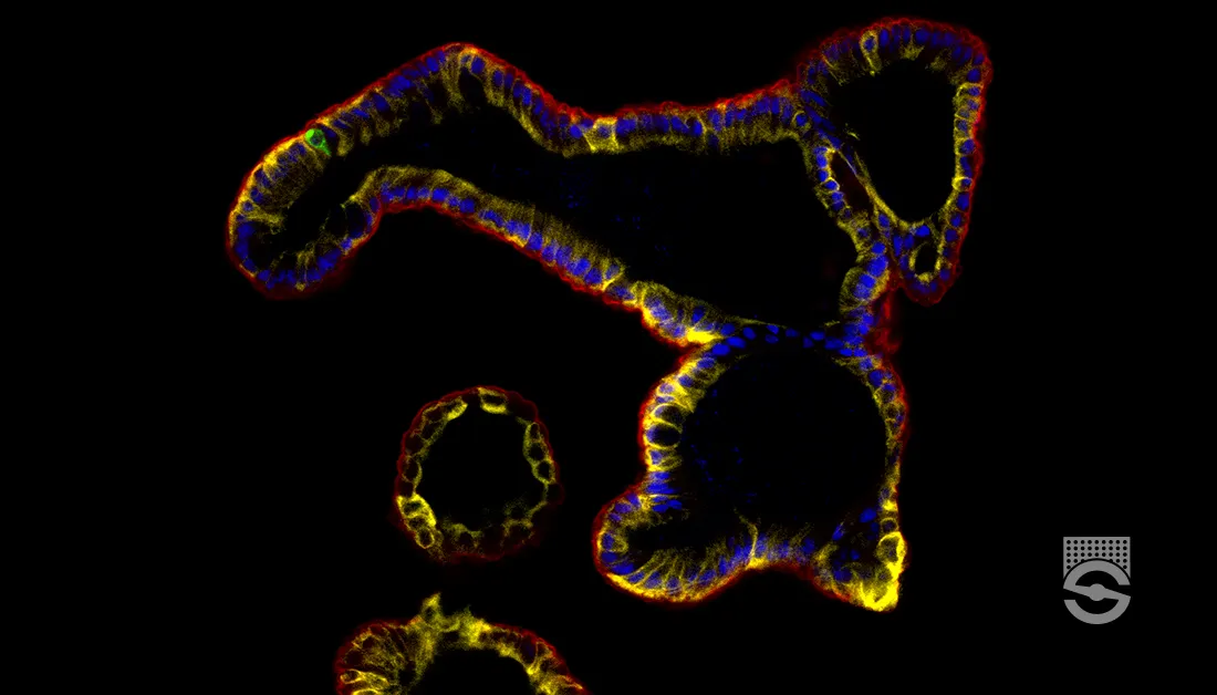
Category

Elastic Fibers

Walter’s Trichrome for Elastic and Collagen

By Dye Type, Elastic Fibers, Orcein, Protocols, Stain Target, Stain Type, Trichrome Staining, Trichrome, One-Step

Walter's Trichrome

for Elastic and Collagen

9
steps
8
materials

Materials

Solution A

MaterialAmount
Ferric ammonium sulphate2.5g
Distilled water100mL

Solution B

MaterialAmount
Phosphotungstic acid2g
Distilled water100mL

Solution C

MaterialAmount
Eosin B0.7g
Acid fuchsin, sat. aqu4mL
Unna’s elastic stain35mL
Ethanol, 50%35mL
Glycerol40mL

Tissue Sample

5µ paraffin sections of neutral buffered formalin fixed tissue are suitable. Other fixatives are likely to be satisfactory. Most trichrome stains benefit from picric acid or mercuric chloride fixation. Formalin fixed tissues may benefit from secondary fixation of sections in Bouin’s fluid.

Protocol

  1. Bring sections to water via xylene and ethanol.
  2. Place into solution A for 24 hours.
  3. Rinse quickly with distilled water.
  4. Place into solution B for 10 minutes.
  5. Rinse quickly with distilled water.
  6. Place into solution C for 15-20 minutes.
  7. Rinse quickly with 95% ethanol until clouds of dye stop being extracted.
  8. Dehydrate with 100% ethanol for 1 minute.
  9. Clear with xylene and mount with a resinous medium.

Expected Results

  • Nuclei  –  purple
  • Elastic fibres  –  purple
  • Erythrocytes  –  orange
  • Collagen  –  blue

Notes

Safety Note

Prior to handling any chemical, consult the Safety Data Sheet (SDS) for proper handling and safety precautions.

References

  1. Gray, Peter. (1954)
    The Microtomist’s Formulary and Guide.
    Originally published by: The Blakiston Co.
    Republished by: Robert E. Krieger Publishing Co.
    Citing:
    Walter, (1930)
    Zeitschrift für wissenschaftliche Mikroskopie und für mikroskipische Technik,
    v. 46, pp. 458

French’s Stain for Elastic Fibres

By Dye Type, Elastic Fibers, Iron Resorcin, Protocols, Stain Target, Stain Type

French's Stain

for Elastic Fibres

7
steps
9
materials

Materials

  • Weigert’s iron hematoxylin or equivalent
  • Van Gieson’s picro-fuchsin
  • French’s solution
    MaterialAmount
    Basic fuchsin1g
    Crystal violet1g
    Resorcin4g
    Ferric chloride, 30% aqu.25mL
    Ethanol 95%200mL
    Distilled water200mL
    Hydrochloric acid, conc.4mL

    Preparation

    1. Add the dye and resorcin to the water in an oversize flask.
    2. Bring to the boil, and add the ferric chloride.
    3. Boil for 5 minutes.
    4. Cool and filter.
    5. Dry the filter paper and beaker. Place the precipitate and filter paper back into the flask.
    6. Add the ethanol and heat carefully until the precipitate dissolves.
    7. Add the hydrochloric acid.
    8. Restore to 200 mL with 95% ethanol.

Tissue Sample

5µ paraffin sections of neutral buffered formalin fixed tissue are suitable. Other fixatives are likely to be satisfactory.

Protocol

  1. Bring sections to water via xylene and ethanol.
  2. Place into French’s solution until adequately stained (may be overnight).
  3. Wash with 95% ethanol to remove excess solution.
  4. Differentiate with 1% acid alcohol.
  5. Wash in water.
  6. Counterstain with iron hematoxylin and van Gieson.
  7. Dehydrate with ethanol, clear with xylene and mount with a resinous medium.

Expected Results

  • Elastic fibres  –  dark blue-green
  • Collagen  –  red
  • Cytoplasm  –  yellow
  • Nuclei  –  blue

Notes

  • This is Weigert’s iron resorcin fuchsin using crystal violet and basic fuchsin instead of basic fuchsin alone.
  • It is usually recommended that the basic fuchsin should not be of the type that produces good Schiff’s reagent. That is, the basic fuchsin should contain more rosanilin than pararosanilin.
  • The ferric chloride solution should be freshly made.

Safety Note

Prior to handling any chemical, consult the Safety Data Sheet (SDS) for proper handling and safety precautions.

References

  1. Histological demonstration techniques, (1974)
    Cook, H C.
    Butterworths, London, England

Weigert’s Stain for Elastic Fibres

By Dye Type, Elastic Fibers, Iron Resorcin, Protocols, Stain Target, Stain Type

Weigert's Stain

for Elastic Fibres

7
steps
8
materials

Materials

  • Weigert’s iron hematoxylin or equivalent
  • Van Gieson’s picro-fuchsin
  • Weigert’s solution
    MaterialAmount
    Basic fuchsin2g
    Resorcin4g
    Ferric chloride, 30% aqu.25mL
    Ethanol 95%200mL
    Distilled water200mL
    Hydrochloric acid, conc.4mL

Preparation

  1. Add the dye and resorcin to the water in an oversize flask. Bring to the boil, and add the ferric chloride.
  2. Boil for 5 minutes.
  3. Cool and filter. Dry the filter paper and beaker. Place the precipitate and filter paper back into the flask.
  4. Add the ethanol and heat carefully until the precipitate dissolves.
  5. Add the hydrochloric acid.
  6. Restore to 200 mL with 95% ethanol.

Tissue Sample

5µ paraffin sections of neutral buffered formalin fixed tissue are suitable. Other fixatives are likely to be satisfactory.

Protocol

  1. Bring sections to water via xylene and ethanol.
  2. Place into Weigert’s solution for 20 minutes to 1 hour.
  3. Wash with 95% ethanol to remove excess solution.
  4. Differentiate with 1% acid alcohol.
  5. Wash in water.
  6. Counterstain with iron hematoxylin and van Gieson.
  7. Dehydrate with ethanol, clear with xylene and mount with a resinous medium.

Expected Results

  • Elastic fibres – blue black
  • Collagen – red
  • Cytoplasm – yellow
  • Nuclei – blue

Notes

  • The procedure often benefits from a Mallory bleach.
  • It is usually recommended that the basic fuchsin should not be of the type that produces good Schiff’s reagent. That is, the basic fuchsin should contain more rosanilin than pararosanilin.
  • The ferric chloride solution should be freshly made.

Safety Note

Prior to handling any chemical, consult the Safety Data Sheet (SDS) for proper handling and safety precautions.

References

  1. Culling C.F.A., (1974)
    Handbook of histopathological and histochemical techniques, Ed. 3
    Butterworth, London, UK.
  2. Drury, R.A.B. and Wallington, E.A., (1980)
    Carleton’s histological technique, Ed. 5
    Oxford University Press, Oxford, UK.
  3. Humason, G. L., (1967)
    Animal tissue techniques, Ed. 2
    W. H. Freeman and Company, San Francisco, Ca, USA

Verhoeff’s Iron Hematoxylin for Elastic

By Dye Type, Elastic Fibers, Hematoxylin and Eosin Staining, Iron Hematoxylin, Mordanted Hematoxylin, Protocols, Stain Target, Stain Type

Verhoeff's Iron Hematoxylin

for Elastic

10
steps
7
materials

Materials

Stock Verhoeff A

MaterialAmount
Hematoxylin1g
Ethanol, absolute20mL

Stock Verhoeff B

MaterialAmount
Ferric chloride10g
Distilled water100mL

Stock Verhoeff C

MaterialAmount
Potassium iodide4g
Iodine2g
Distilled water100mL

Working solution

MaterialAmount
Stock solution A20mL
Stock solution B8mL
Stock solution C8mL

Differentiator

MaterialAmount
Stock solution B10mL
Distilled water40mL

Tissue Sample

A 10% Formalin variant is suitable. Paraffin sections at 5µ are preferred.

Protocol

  1. Bring sections to water via xylene and ethanol.
  2. Place in working Verhoeff’s solution for 15 minutes.
  3. Wash well with tap water to remove all excess hematoxylin.
  4. Differentiate until the elastic fibres are satisfactory. This should be controlled microscopically based on the appearance of fibres in the area of interest.
  5. Rinse well with tap water.
  6. Place into 95% ethanol for 5 minutes to remove iodine discolouration.
  7. Wash well with tap water to remove all residual chemicals.
  8. Counterstain with Van Gieson.
  9. Dehydrate with absolute ethanol.
  10. Clear with xylene and mount with a resinous medium

Expected Results

  • Nuclei  –  black
  • Elastic fibres  –  black
  • Muscle  –  yellow
  • Collagen  –  red

Notes

  • The alcoholic hematoxylin solution should be freshly made. It does not need to have been ripened as ferric chloride is an oxidising agent. Some technologists keep pre-weighed aliquots of 1 gram hematoxylin and dissolve it in 20 mL ethanol as needed.
  • The working solution should be made just before use and allowed to stand for five minutes to ripen.
  • While this is a popular elastic stain, it is difficult to stain both coarse fibres and fine fibres optimally in a single section. If coarse fibres are well stained, the finest fibres may be completely decolourised, and if fine fibres are optimally stained, the coarse fibres are underdifferentiated. Ensure differentiation is optimised for the fibres of interest.
  • If Van Gieson is used as a counterstain, the elastic fibres should be very slightly underdifferentiated as picric acid will also remove some hematoxylin staining.
  • This method likely stains by two mechanisms: ionic attachment of iron mordanted hematoxylin to most structures and by van der Waal’s forces to elastic fibres. The differentiation step is accomplished either by oxidative bleaching of hematoxylin by ferric chloride, or by displacement of the mordanted hematoxylin by free mordant in solution. The dye attached to elastic fibres by van der Waal’s forces is not extracted easily. It is, however, still susceptible to bleaching, which likely explains why fine fibres may be pale. That bleaching takes place is shown by the differentiating fluid remaining clear during differentiation, extracted dye being seen to bleach as it dissolves out.
  • Note that nuclei are only adequately, not well, stained.

Safety Note

Prior to handling any chemical, consult the Safety Data Sheet (SDS) for proper handling and safety precautions.

References

  1. Histological demonstration techniques, (1974)
    Cook, H C.
    Butterworths, London, England
  2. Culling, C.F.A., (1963)
    Handbook of histopathological techniques, 2nd ed.
    Butterworths, London.

Unna’s Orcein-Aniline Blue for Elastic

By Dye Type, Elastic Fibers, Orcein, Protocols, Stain Target, Stain Type

Unna's Orcein-Aniline Blue

for Elastic

7
steps
7
materials

Materials

  • A red nuclear stain
  • Solution A
    MaterialAmount
    Acetic acid, glacial1mL
    Distilled water1L
  • Solution B
    MaterialAmount
    Aniline blue0.5g
    Orcein0.5g
    Acetic acid, glacial2.5mL
    Ethanol, 100%25mL
    Distilled water50mL
    Glycerol10mL

Preparation

  1. Dissolve the aniline blue in the water with heat.
  2. Cool and filter.
  3. Dissolve the orcein in the ethanol.
  4. Add the acetic acid and glycerol.
  5. Combine the solutions.

Tissue Sample

5µ paraffin sections of neutral buffered formalin fixed tissue are suitable. Other fixatives are likely to be satisfactory.

Protocol

  1. Bring sections to water via xylene and ethanol.
  2. Stain the nuclei with a red nuclear stain.
  3. Place into solution A for a few minutes.
  4. Place into solution B for 1-10 hours.
  5. Differentiate with solution A until elastic fibres are clear.
  6. Dehydrate with 95% and absolute ethanols.
  7. Clear with xylene and mount with a resinous medium.

Expected Results

  • Nuclei  –  red
  • Elastic  –  reddish brown
  • Bone  –  reddish brown
  • Background  –  blue

Notes

  • Gatenby & Beams specified water blue rather than aniline blue
  • Unna’s method has been more commonly applied in conjunction with other methods, such as Pasini’s trichrome, with solution B (Unna’s solution) being incorporated into other staining solutions.

Safety Note

Prior to handling any chemical, consult the Safety Data Sheet (SDS) for proper handling and safety precautions.

References

  1. Gray, Peter. (1954)
    The Microtomist’s Formulary and Guide.
    Originally published by: The Blakiston Co.
    Republished by: Robert E. Krieger Publishing Co.
    Citing:
    Gatenby, J. B. & Cowdry, E. V., (1928)
    Bolles Lee’s Microtomist’s Vade Mecum, pp. 280
    Blakiston, Philadelphia

Taenzer-Unna Orcein for Elastic Fibres

By Dye Type, Elastic Fibers, Orcein, Protocols, Stain Target, Stain Type

Taenzer-Unna Orcein

for Elastic Fibres

8
steps
3
materials

Materials

Orcein solution

MaterialAmount
Orcein1g
Ethanol, 70%100mL
Hydrochloric acid, conc.1mL

Acid ethanol

MaterialAmount
Ethanol, 70%100mL
Hydrochloric acid, conc.1mL

Tissue Sample

5µ paraffin sections of neutral buffered formalin fixed tissue are suitable. Other fixatives are likely to be satisfactory.

Protocol

  1. Bring sections to water via xylene and ethanol.
  2. Rinse with 70% ethanol.
  3. Place into orcein solution for an appropriate time.
  4. Optionally, treat with acid ethanol until collagen is unstained (a few seconds).
  5. Wash with water
  6. Counterstain if wished.
  7. Dehydrate with 95% and absolute ethanols.
  8. Clear with xylene and mount with a resinous medium.

Expected Results

  • Elastic fibres  –  purplish brown
  • Other tisse  –  as counterstained

Notes

  • Gray specifies 0.8 grams orcein, most others specify 1 gram.
  • Gray specifies 50% ethanol, Culling 70% and Carleton 80%. Others have recommended concentrations ranging from 65% to 100% (Lillie).
  • Staining times specified include 6-12 hours (Gray), overnight (Culling) and 30 minutes to 2 hours (Carleton).
  • Elevating the temperature (37°C Carleton, 56°C Culling) decreases staining time.
  • Recommended counterstains are van Gieson, methylene blue, H&E.

Safety Note

Prior to handling any chemical, consult the Safety Data Sheet (SDS) for proper handling and safety precautions.

References

  1. Gray, Peter. (1954)
    The Microtomist’s Formulary and Guide.
    Originally published by: The Blakiston Co.
    Republished by: Robert E. Krieger Publishing Co.
    Citing:
    von Kahlden, C., and Laurent, O. (1896)
    Technique microscopique.
    Carré, Paris, France
  2. Drury, R.A.B. and Wallington, E.A., (1980)
    Carleton’s histological technique Ed. 5
    Oxford University Press, Oxford, UK.
  3. Culling C.F.A., (1974)
    Handbook of histopathological and histochemical techniques Ed. 3
    Butterworth, London, UK.
  4. Mallory, F. B. & Wright, J.H., (1904)
    Pathological technique, Ed.3
    W. B. Saunders, Philadelphia, USA.
  5. Lillie, R.D., (1954)
    Histopathologic technique and practical histochemistry Ed.2
    Blakiston, New York, USA.

Sheridan’s Stain for Elastic Fibres

By Dye Type, Elastic Fibers, Iron Resorcin, Protocols, Stain Target, Stain Type

Sheridan's Stain

for Elastic Fibres

7
steps
9
materials

Materials

  • Weigert’s iron hematoxylin or equivalent
  • Van Gieson’s picro-fuchsin
  • Sheridan’s solution
    MaterialAmount
    Crystal violet2g
    Dextrin2g
    Resorcin4g
    Ferric chloride, 30% aqu.25mL
    Ethanol 95%200mL
    Distilled water200mL
    Hydrochloric acid, conc.4mL

Preparation

  1. Add the dye, dextrin and resorcin to the water in an oversize flask. Bring to the boil, and add the ferric chloride.
  2. Boil for 5 minutes.
  3. Cool and filter.
  4. Dry the filter paper and beaker. Place the precipitate and filter paper back into the flask.
  5. Add the ethanol and heat carefully until the precipitate dissolves.
  6. Add the hydrochloric acid.
  7. Restore to 200 mL with 95% ethanol.

Tissue Sample

5µ paraffin sections of neutral buffered formalin fixed tissue are suitable. Other fixatives are likely to be satisfactory.

Protocol

  1. Bring sections to water via xylene and ethanol.
  2. Place into Sheridan’s solution until adequately stained (may be overnight)).
  3. Wash with 95% ethanol to remove excess solution.
  4. Differentiate with 1% acid alcohol.
  5. Wash in water.
  6. Counterstain with iron hematoxylin and van Gieson.
  7. Dehydrate with ethanol, clear with xylene and mount with a resinous medium.

Expected Results

  • Elastic fibres  –  green
  • Collagen  –  red
  • Cytoplasm  –  yellow
  • Nuclei  –  blue

Notes

  • This is similar to Weigert’s iron resorcin fuchsin, but substitutes crystal violet and dextrin for basic fuchsin.
  • The ferric chloride solution should be freshly made.

Safety Note

Prior to handling any chemical, consult the Safety Data Sheet (SDS) for proper handling and safety precautions.

References

  1. Histological demonstration techniques, (1974)
    Cook, H C.
    Butterworths, London, England

Musto’s Hematoxylin for Elastic Fibres

By Dye Type, Elastic Fibers, Iron Hematoxylin, Protocols, Stain Target, Stain Type

Musto's Hematoxylin

for Elastic Fibres

10
steps
11
materials

Materials

Stock hematoxylin

MaterialAmount
Hematoxylin2g
Ethanol, 95%100mL

Stock ferric chloride

MaterialAmount
Ferric chloride, hexahydrate12.4g
Distilled water500mL
Hydrochloric acid, concentrated5mL

Stock iodine

MaterialAmount
Potassium iodide4g
Iodine2g
Distilled water100mL

Thiosulphate solution

MaterialAmount
Sodium thiosulphate5g
Distilled water100mL

Working solution

MaterialAmount
Stock hematoxylin30mL
Stock ferric chloride20mL
Stock iodine10mL

Tissue Sample

A 10% Formalin variant is suitable. Paraffin sections at 5µ are preferred.

Protocol

  1. Bring sections to water via xylene and ethanol.
  2. Place sections in Bouin’s fluid at 60°C for 1 hour.
  3. Remove the yellow colouration with running tap water.
  4. Place in working solution for 45 minutes.
  5. Wash well with tap water, then rinse with distilled water.
  6. Place into thiosulphate solution for 2 minutes to remove iodine discolouration.
  7. Wash well with tap water, then rinse with distilled water.
  8. Counterstain with Van Gieson.
  9. Dehydrate with absolute ethanol.
  10. Clear with xylene and mount with a resinous medium

Expected Results

  • Nuclei – black
  • Elastic fibres – black
  • Muscle – yellow
  • Collagen – red

Notes

  • The original paper recommended counterstaining with a modification of the HPS.
  • The treatment with Bouin’s solution is likely included to improve an HPS stain. It may be possible to eliminate it for counterstaining with Van Gieson.
  • This is a modification of Verhöeff’s hematoxylin which does not require differentiating, although the Van Gieson counterstain may diminish the intensity of the stain.

Safety Note

Prior to handling any chemical, consult the Safety Data Sheet (SDS) for proper handling and safety precautions.

References

  1. Musto, Linda, (1981),
    Improved iron-hematoxylin stain for elastic fibers.,
    Stain Technology, v 56, page 185-7.

Musto’s Hematoxylin Scarlet-Saffron for Elastic

By Dye Type, Elastic Fibers, Iron Hematoxylin, Protocols, Stain Target, Stain Type

Musto's Hematoxylin Scarlet-Saffron

for Elastic

14
steps
16
materials

Materials

Stock hematoxylin

MaterialAmount
Hematoxylin2g
Ethanol, 95%100mL

Stock ferric chloride

MaterialAmount
Ferric chloride, hexahydrate12.4g
Distilled water500mL
Hydrochloric acid, concentrated5mL

Stock iodine

MaterialAmount
Potassium iodide4g
Iodine2g
Distilled water100mL

Working solution

MaterialAmount
Stock hematoxylin30mL
Stock ferric chloride20mL
Stock iodine10mL

Tungstophosphoric acid

MaterialAmount
Tungstophosphoric acid5g
Distilled water100mL

Acetic acid

MaterialAmount
Acetic acid, glacial1mL
Distilled water99mL

Woodstain scarlet

MaterialAmount
Woodstain scarlet, 1% aqu.50mL
Tungstophosphoric acid2g

Prepare fresh.

Alcoholic saffron solution

MaterialAmount
Saffron1g
Ethanol, absolute100mL

Extract the saffron in ethanol at 56°C for 24 hours, shaking occasionally. Cool and filter.

Tissue Sample

A 10% Formalin variant is suitable. Paraffin sections at 5µ are preferred.

Protocol

  1. Bring sections to water via xylene and ethanol.
  2. Place sections in Bouin’s fluid at 60°C for 1 hour.
  3. Remove the yellow colouration with running tap water.
  4. Place in working solution for 45 minutes.
  5. Wash well with tap water, then rinse with distilled water.
  6. Place into the woodstain scarlet solution for 5 minutes.
  7. Rinse well with distilled water.
  8. Differentiate in tungstophosphoric acid solution for 5 minutes.
  9. Transfer directly to acetic acid solution for 5 minutes.
  10. Dehydrate with 95% ethanol for 1 minute.
  11. Place in absolute ethanol, two changes, for 1 minute each.
  12. Place in alcoholic saffron solution for 5-10 minutes.
  13. Place in absolute ethanol, two changes, for 1 minute each.
  14. Clear with xylene and mount with a resinous medium

Expected Results

  • Nuclei – black
  • Elastic fibres – black
  • Muscle – pale red
  • Collagen – yellow

Notes

Safety Note

Prior to handling any chemical, consult the Safety Data Sheet (SDS) for proper handling and safety precautions.

References

  1. Musto, Linda, (1981),
    Improved iron-hematoxylin stain for elastic fibers.,
    Stain Technology, v 56, page 185-7.

Miller’s Stain for Elastic Fibres

By Dye Type, Elastic Fibers, Iron Resorcin, Protocols, Stain Target, Stain Type

Miller's Stain

for Elastic Fibres

8
steps
12
materials

Materials

Preparation

  1. In an oversized flask heat the water and add the three dyes.
  2. When dissolved, add the resorcin, dextrin and ferric chloride in the order given.
  3. Boil for 5 minutes, then filter while hot.
  4. Place the precipitate and filter paper back into the original flask, add the 95% ethanol and simmer gently for 20 minutes.
  5. Cool and filter.
  6. Restore the volume to 200 mL with 95% ethanol, then add 2 mL of concentrated hydrochloric acid.

Tissue Sample

5µ paraffin sections of neutral buffered formalin fixed tissue are suitable. Other fixatives are likely to be satisfactory.

Protocol

  1. Bring sections to water via xylene and ethanol.
  2. Do a Mallory bleach with 0.25% potassium permanganate for ten minutes.
  3. Rinse with 95% ethanol.
  4. Place into Miller’s solution up to three hours.
  5. Wash with 95% ethanol to remove excess solution.
  6. Wash in water.
  7. Counterstain with iron hematoxylin and van Gieson.
  8. Dehydrate with ethanol, clear with xylene and mount with a resinous medium.

Expected Results

  • Elastic fibres  –  black
  • Nuclei  –  black
  • Cytoplasm  –  yellow
  • Collagen  –  red

Notes

  • Three hours is recommended, although staining may be adequate after 90 minutes.
  • Dilute with an equal quantity of 95% ethanol for overnight staining.
  • The staining solution is stable for years.

Safety Note

Prior to handling any chemical, consult the Safety Data Sheet (SDS) for proper handling and safety precautions.

References

  1. Miller, P.J., (1971)
    Journal of Medical Laboratory Technology, V. 28
  2. Ellis, R.,
    Miller’s Elastic Staining Protocol
    IHC World